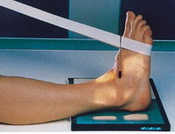
IR Size: 8x10 TT
SID: 40''
• Pt. Supine with Leg Extended
• Foot dorsiflexed 90°
• _|_ between malleoli
•
•
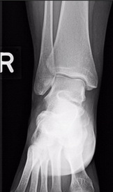
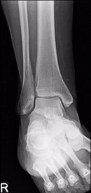
• Open Tibiotalar Joint and centered
• Normal overlap of tibiofibular joint with anterior tubercle slightly SI over the fibula
• No SI of medial talomalleolar joint
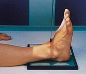
IR Size: 8x10 TT
SID: 40''
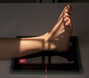
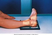
• Pt. Supine w/ Leg Extended, Foot 90°
• For tibiofibular joint in profile - Leg & Foot 45° Medially
• For Mortise Joint (99% of the time) - Leg and foot 15°-20° Medially
• _|_ between malleoli, For Mortise Joint - Malleoli Parallel
•
•
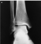
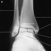
• Mortise Joint or Tibiofibular Joint
• For mortise - Complete open mortise joint with tibiotalar joint space in profile
• For 45° - Some overlap of tibia & fibula over the talus
• Open tibiofibular joint
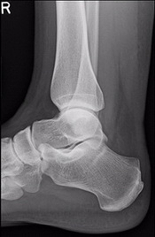
IR Size: 8x10 TT
SID: 40''
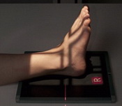
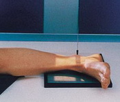
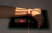
• Pt lateral position on affected
• Foot dorsiflexed 90°
• (Usually Knee should be off the table, use sponge)
• _|_ on medial malleolus
•
• Include 5th MT tuberosity in case of Jones Fx
•
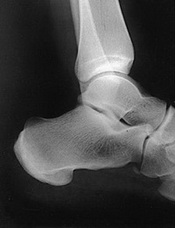
• Tibiotalar joint well seen and centered with SI of medial & lateral talar domes
• Fibula over posterior half of tibia
• 5th MT seen to check for Jones fx
IR Size: 8x10 TT
SID: 40''
• Same as AP & AP oblique
• Uses Extreme Inversion & Eversion
• If ligament is torn a widening of the joint space is demonstrated one of injury
• Radiologist performs procedure
•
•
•
• Ligamentous Tears
